Minimally invasive

Lumbar spine stabilization surgery

When abnormal motion between the vertebrae (instability) is the source of pain, stabilization surgery may be required. Several techniques are available — the choice is always case-dependent. My deepest experience is in the MI-TLIF technique, which is less invasive and achieves lasting stability while preserving the surrounding muscles.

Jump to When it is needed First step: non-surgical MI-TLIF Other techniques What to expect Patient case FAQ

When is stabilization needed?

As lumbar discs wear and age advances, the connection between the vertebrae can weaken. This leads to abnormal hypermobility — instability — whose most characteristic symptom is recurring "locking" lower back pain after physical work or load. In more severe cases, vertebral slippage (spondylolisthesis) can develop.

The aim of surgery is to place an internal fixation and a bone-graft substitute, allowing the two vertebrae to fuse over the long term. Once fusion has occurred, the internal fixation no longer plays an active role — but it does not need to be removed.

Indication for urgent surgery

  • Pain syndrome unresponsive to medication
  • Lower limb paralysis — muscle weakness
  • Cauda equina syndrome — bowel and bladder dysfunction (medical emergency)

Indication for planned surgery

  • Recurring lower back pain unresponsive to non-surgical treatment
  • Significant instability or vertebral slippage confirmed by imaging (MRI, CT, dynamic X-ray)
  • Persistent symptoms that impair quality of life and work
  • Persistent nerve compression with stenosis combined with instability

Non-surgical treatment — where possible

Stabilization surgery is a major intervention with a long recovery. The surgical route is justified only when non-surgical options have been exhausted — or when the clinical picture clearly requires surgery. Most instability complaints can also improve as a first step with targeted treatment and physiotherapy.

The non-surgical path is worth trying

Targeted CT-guided block injections, infusion therapy and stabilizing physiotherapy — always worth exploring thoroughly before surgery. Details on the non-surgical treatment page.

More on non-surgical treatment

MI-TLIF — minimally invasive stabilization

The current gold standard is open TLIF — a reliable, classical technique proven over decades. MI-TLIF developed as a less invasive variant: instead of detaching the muscles, we work through them with percutaneous screws. Clinical results are similar, but the patient experience is significantly better.

Minimally invasive spine stabilization surgery — intraoperative image
MI-TLIF surgery: percutaneous screw placement and exposure through a tubular retractor — preserving the surrounding muscles

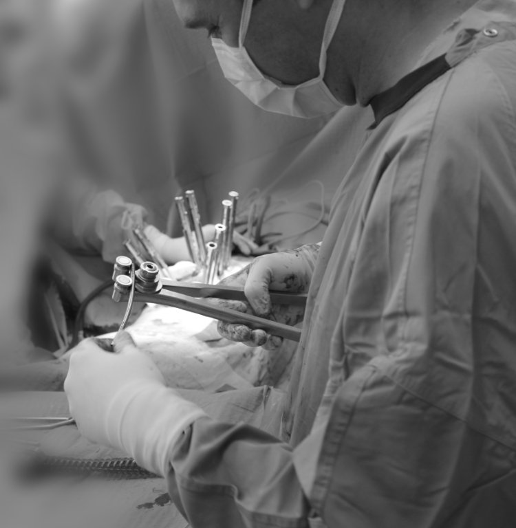
The essence of the technique: percutaneous titanium screws are placed into the pedicles of the vertebrae through small skin incisions, under imaging guidance. Between two adjacent screws, a tube (tubular retractor) is advanced down to the spine, splitting the muscle fibres apart — not detaching them. Through this corridor, the damaged disc is removed and a cage filled with bone-graft substitute is placed. The screws are connected with rods, securing stability.

Muscle preservation

Muscle fibres are not detached from the bone — they are split apart. The function of the surrounding musculature is better preserved, which is particularly advantageous for younger, active patients.

Less blood loss

Minimal exposure and targeted work significantly reduce intraoperative blood loss compared to the open technique.

Shorter hospital stay

Faster mobilization and less postoperative pain typically translate into a shorter hospital stay.

Faster recovery

Muscle preservation and reduced tissue trauma allow a faster return to everyday activity, with less postoperative muscle pain.

Note: MI-TLIF is technically more demanding than classical open TLIF — which is why fewer surgeons perform it routinely. Beyond the long learning curve, it requires specialised imaging equipment and instrumentation. This is the technique in which I have built the deepest experience over the years, and for younger patients I typically choose this approach.

The full repertoire

Several techniques are established in lumbar stabilization. Each has its own advantages and trade-offs — the choice is determined by the individual case: the patient's condition, lifestyle, the location of the segment to be operated on, and the type of pathology. In addition to MI-TLIF, I perform the following techniques (with the exception of XLIF), so we can discuss together at the consultation which one best fits your situation.

TLIF Open transforaminal lumbar interbody fusion

The classical, current gold standard procedure. Longer skin incision, with the muscles detached from the bone — leading to a longer recovery and more postoperative muscle pain, but reliable, predictable results. Applicable across the full spectrum of instability.

OLIF Oblique lateral interbody fusion

The spine is reached through an oblique anterior approach. Its advantage is the ability to insert a larger cage; its disadvantage is that it is usually combined with posterior percutaneous stabilization (more incisions, longer surgery). At certain levels and in higher-grade deformities it offers a clear benefit.

ALIF Anterior lumbar interbody fusion

The spine is approached from the front through the abdomen. It allows the largest cage to be placed, but its sensitivity — proximity to large vessels — requires special care. Mainly considered at the lower lumbar segments.

XLIF Lateral interbody fusion

Approach from the side, through the psoas muscle. It works through a narrow anatomical "window" and requires specialised instrumentation. I do not perform this technique personally — if XLIF were indicated, I would refer the patient to my colleague at the institution.

The logic behind the decision: selecting a technique is not a "competition" between methods, but a case-dependent clinical decision. Based on MRI, CT, dynamic X-ray and physical examination, we choose the best solution together at the consultation. For younger, active patients I always weigh the muscle-sparing approach — which is why MI-TLIF is often the choice.

Realistic expectations

Stabilization surgery is a major intervention that can have excellent long-term outcomes — but it is worth being clear in advance about realistic timelines. The postoperative period spans not weeks but months until full recovery is reached.

What to expect

  • Gradual mobilization and walking with support in the days after surgery
  • Hospital stay: typically 3–5 days
  • Postoperative pain decreases significantly over 2–4 weeks
  • Return to office work after 4–6 weeks
  • Physical work: 3–6 months of recovery recommended
  • Lasting freedom from symptoms long-term, if fusion progresses well

What to be prepared for

  • Vertebral fusion takes 6–12 months
  • Full recovery spans 6–12 months, with verification of fusion
  • Regular physiotherapy in the postoperative period — not optional, but a precondition for success
  • CT follow-up at 6 and 12 months to assess fusion
  • Implanted screws and cage remain permanently in the body
  • Adjacent segment syndrome — risk of involvement of a neighbouring level years later

A typical MI-TLIF patient journey

The composite case below shows how a 48-year-old patient working in physical labour moved from "locking" lower back pain through surgical treatment to a full return to work.

MI-TLIF · physical worker

"My back was locking up more and more often"

A 48-year-old man working in physical labour, with painful episodes increasing in frequency over years. MRI showed significant disc wear combined with instability. Non-surgical treatment provided only temporary relief. After MI-TLIF surgery, gradual mobilization and targeted physiotherapy — within a few months the patient returned to physical work.

Read full case

What patients most often ask

Why MI-TLIF, and how does it differ from open TLIF?

Open TLIF is currently the gold standard for lumbar fusion surgery — a reliable technique proven over decades. MI-TLIF achieves the same outcome in a less invasive way: instead of detaching the muscles, we work through them, using smaller incisions and percutaneous screws.

Clinical outcomes — pain reduction and vertebral fusion — are similar between the two techniques. However, muscle preservation results in less blood loss, a shorter hospital stay, less postoperative muscle pain, and faster recovery. It is particularly worth choosing for younger, active patients.

How long does it take for the vertebrae to fuse?

Fusion typically develops over 6–12 months. Bone formation is verified by CT at 6- and 12-month follow-ups. Complete bony remodelling can take additional months.

The implanted titanium screws and the cage remain permanently in the body — removal is not necessary, since once fusion has occurred their role is passive.

How long is the hospital stay?

For MI-TLIF, typically 3–5 days. The patient begins walking with support the day after surgery — an important part of the rapid recovery. The conditions for discharge are independent mobilization and adequate pain control.

When can I return to work?

Return to office work is possible after 4–6 weeks, gradually. For physical work, 3–6 months are recommended, depending on the nature of the work and the progress of fusion. For heavy physical work, returning is appropriate only after fusion is verified — assessed by follow-up CT.

Driving is typically recommended after 4–6 weeks, when painkillers can be discontinued and reflexes have recovered.

How does physiotherapy help after surgery?

Physiotherapy is one of the most important elements of recovery — not an option. In the postoperative period, gradual strengthening of the deep stabilising muscles is essential so that the spine remains stable around the fused segment over the long term.

The programme is typically progressive: breathing exercises and gentle mobilization in the first weeks, followed by increasing core stabilization exercises. I work together with the physiotherapy team at Budai Egészségközpont.

What is the risk of another segment becoming affected later?

The segments above and below the fused level take on increased load — this is known as adjacent segment syndrome. The risk emerges years later and is proportional to the number of fused segments. With single-level fusion, this risk is lower.

Regular physiotherapy, good posture and weight control help prevent it. If new symptoms arise later, we evaluate them with follow-up imaging.

What is most important in preparation for surgery?

Preoperative physiotherapy — bringing the muscles into the best possible condition — significantly aids recovery. Stopping smoking (ideally 6–8 weeks before surgery) is proven to improve the chances of successful fusion. Modification of any anticoagulant medication is handled individually and discussed at the consultation.

Dr. Zsolt Szövérfi
Written and medically reviewed by Dr. Zsolt Szövérfi PhD Spine Surgeon · Orthopaedic and Trauma Specialist · Full profile Last updated: April 2026

Book a consultation

If spine stabilization has come up as a possibility, the first step is a thorough spine surgical consultation — reviewing the existing imaging and discussing the appropriate technique together.

Book Appointment
Second-opinion consultation