Treatment of disc herniation

Around 70% of disc herniations heal without surgery, with conservative treatment. Dr. Zsolt Szövérfi PhD, spine surgeon (Budai Egészségközpont, Budapest), builds on precise diagnosis and targeted non-surgical care; minimally invasive surgery is considered only when symptoms persist or nerve damage is present.

~70% resolve with non-surgical treatment
Jump to What is disc herniation First step: non-surgical Lumbar herniation Cervical herniation Patient cases

What is disc herniation?

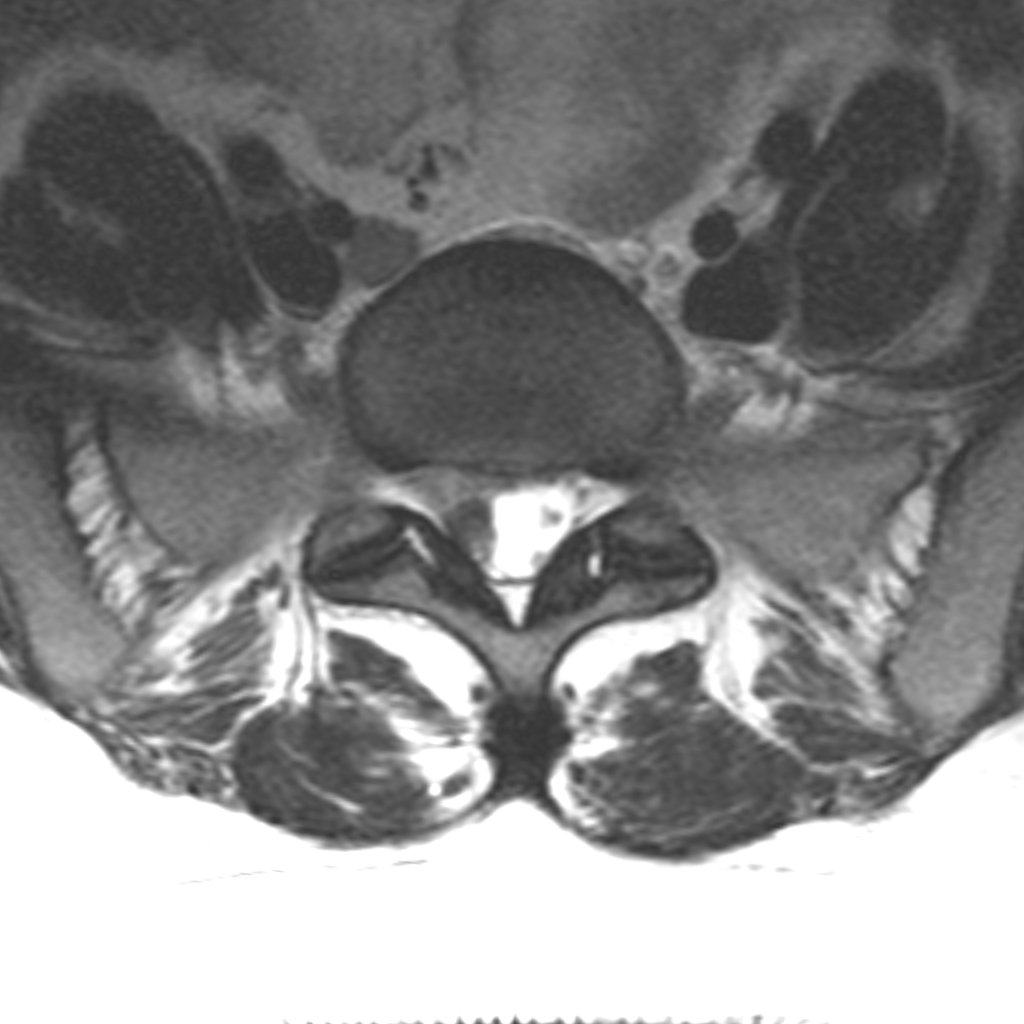
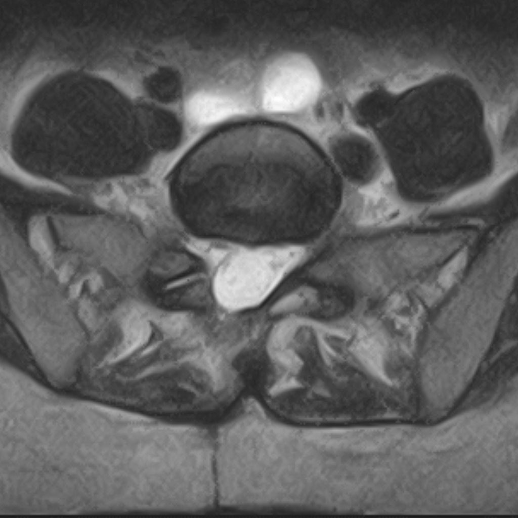
The intervertebral discs between the vertebrae act as flexible cushions that absorb load and allow movement. The fibrous outer wall of the disc (annulus fibrosus) can weaken over time, and a bulge can form on it, putting pressure on the nerves running directly alongside.

Another presentation is the sequestrated disc herniation: a tear forms in the fibrous ring, through which the gel-like material from inside the disc (nucleus pulposus) escapes outside the disc. In this case, in addition to the mechanical pressure on the nerve, the chemical properties of the displaced material also irritate the nerves. Pressure and chemical irritation together create inflammation in the neural elements, which sustains the pain syndrome.

Axial MRI image — lumbar disc herniation
MRI: lumbar disc herniation — disc material compressing the nerve root
Axial MRI image — postoperative state
MRI: postoperative state — the nerve root is decompressed

A herniation on the scan does not, by itself, mean surgery

Spinal imaging frequently shows disc changes even in people who have no symptoms at all. Large studies of symptom-free individuals find that close to half of those around age 40 have some form of disc bulge — without it causing any symptoms. This proportion rises further with age.

This has an important consequence for the patient: the words “disc herniation” on a report do not, on their own, justify surgery. The treatment decision always rests on the symptoms, the physical examination and the scan considered together — not on the image alone. This is why, even alongside an alarming-sounding MRI report, non-surgical treatment is often the right path.

Non-surgical treatment — sufficient in most cases

If disc herniation is suspected, an MRI examination is needed for accurate diagnosis. If the clinical picture does not suggest urgent surgical indication, the first step is almost always non-surgical treatment. A significant proportion of disc herniations resolve durably within a few weeks to months with targeted injection therapy, anti-inflammatory medication and physiotherapy — without surgical intervention.

~70%

Resolves without surgery

A significant proportion of patients with disc herniation become lastingly symptom-free without surgery. A detailed overview of non-surgical treatment is available on the dedicated page.

More on non-surgical treatment

Why is improvement expected without surgery?

In disc herniation, the body has a natural capacity to heal. The protruding or extruded disc material is, over time, substantially reabsorbed by the body — studies show that in roughly two-thirds of cases the herniation measurably shrinks on imaging over the following months. This biological process explains why symptoms ease in many patients without any intervention.

It is important, however, to distinguish two things: the reabsorption visible on a scan and actual freedom from symptoms are not the same. Clinical improvement — the resolution of pain and numbness — often occurs sooner than any visible shrinkage on imaging, and the two do not always go together. The goal of treatment is therefore always to improve symptoms and quality of life, not to “fix” the scan.

What does a disc herniation on an MRI mean? →

Lumbar disc herniation

Disc herniation in the lower back typically causes pain radiating into the leg (sciatica), often with numbness and muscle weakness. The pattern of symptoms helps to identify the responsible segment — but accurate diagnosis requires MRI.

Sciatica — leg pain that radiates: causes and treatment →

Indication for urgent surgery

  • Intolerable pain unresponsive to medication
  • Lower limb paralysis — muscle weakness, difficulty lifting the foot
  • Cauda equina syndrome — bowel and bladder dysfunction, a medical emergency

Indication for planned surgery

  • 6–12 weeks of non-surgical treatment have not produced lasting improvement
  • Symptoms persistently impair quality of life and ability to work
  • The size of the herniation on MRI and the clinical picture jointly justify surgery

How much better is surgery, if it does become necessary?

If conservative treatment fails to bring lasting improvement after several weeks, surgery is a reliable solution. Large international studies show that in appropriately selected patients, surgery brings faster pain relief and a faster return to daily life than continued conservative care — and this advantage remains measurable years after the procedure.

This does not mean, however, that surgery is urgent. In the absence of red flags, the non-surgical path can be followed safely: a delay does not worsen the outcome of surgery performed later. The decision is therefore unhurried, and is always made together with the patient — which is exactly why it is worth turning to a surgeon who explores the non-surgical options before recommending an intervention.

Surgical technique: discectomy

The surgery exposes the spine through a small skin incision (2–3 cm), and a minimal bony window is opened in the gap between two vertebrae. Through this corridor, the herniated disc fragment compressing the nerve is removed and the neural elements are decompressed.

Among the techniques currently in use — conventional exposure, microscopic technique (microdiscectomy), endoscopic discectomy — experience suggests there is no clinically significant difference in long-term outcomes. Wherever possible, I always favour the least invasive solution.

Cervical disc herniation

Disc herniation in the cervical spine can cause nerve root or spinal cord compression. Symptoms typically include pain radiating into the arm, numbness, clumsiness in fine motor movements, and in more severe cases gait disturbance or hand weakness.

Indication for urgent surgery

  • Pain unresponsive to medication
  • Upper and/or lower limb paralysis
  • Clinical signs of spinal cord compression (myelopathy) — gait disturbance, loss of fine motor control

Indication for planned surgery

  • Persistent symptoms unresponsive to non-surgical treatment
  • Gradual deterioration
  • MRI morphology justifies surgery (significant herniation, spinal stenosis)

Surgical technique: ACDF — Anterior Cervical Discectomy and Fusion

Through a 3–4 cm skin incision in the front of the neck, the spine is exposed, the disc and the herniation that has extruded into the spinal canal are removed, and the neural elements are decompressed.

A cage is placed in the position of the disc, secured with screws or a plate. The cage contains bone-graft substitute, which allows the neighbouring vertebrae to fuse over time. The procedure can be performed on up to four adjacent segments. In some cases, a mobile disc prosthesis can also be implanted, where preserving the motion of adjacent segments is advantageous.

ACDF relieves symptoms with a high success rate. Recovery time is typically about one month.

Three typical patient journeys

The same diagnosis — three different paths. The composite cases below show how disc herniation treatment is always an individual decision: the non-surgical success, the planned surgery and the urgent intervention are all real-world scenarios.

Non-surgical success

"The pain was already radiating into my leg"

A 40-year-old patient working in an office. After a CT-guided injection and physiotherapy, the patient became lastingly symptom-free — without surgery.

Read full case
Planned surgery

"I could barely walk"

A 45-year-old active man. Non-surgical treatment brought only temporary improvement — after surgery, immediate symptomatic improvement and full recovery.

Read full case
Urgent surgery

"I couldn't use my hand properly"

A 42-year-old dentist with cervical disc herniation and developed paralysis. ACDF surgery within 24 hours, full functional recovery within 3 months.

Read full case
Dr. Zsolt Szövérfi
Written and medically reviewed by Dr. Zsolt Szövérfi PhD Spine Surgeon · Orthopaedic and Trauma Specialist · Full profile Last updated: June 2026

Book a consultation

Whether you have suspected disc herniation or a confirmed diagnosis and want to learn about the treatment options, the first step is a spine surgical consultation.

Book Appointment
Second-opinion consultation
Medically reviewed by: Dr. Zsolt Szövérfi PhD, spine surgeon · Last updated: June 2026