When is Stabilisation Needed?
Degeneration of the lumbar discs and the resulting abnormal motion (instability) can lead to overgrowth of the tissues connecting the vertebrae. When intervertebral instability rather than stenosis is the source of symptoms, stabilisation techniques may be required.
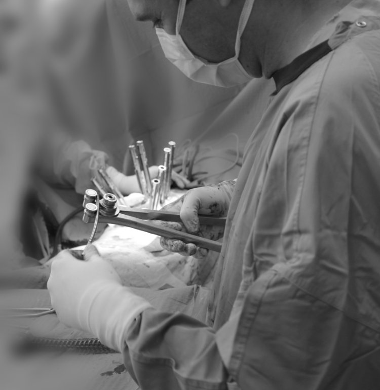
The essence of the surgery is temporary internal fixation and bone graft placement. After bony fusion is achieved, the internal fixation no longer has an active role but does not need to be removed.
Indications for Urgent Surgery
- Intractable pain unresponsive to medication
- Lower extremity paralysis
- Cauda equina syndrome — bowel and bladder dysfunction
Indications for Elective Surgery
- Failure of conservative treatment
- Chronic pain syndrome
- Significant instability on imaging (CT, MRI, X-ray)
Traditional vs. Minimally Invasive Approach
Traditional Open TLIF
- 10–15 cm skin incision
- Stripping of muscles from the spine
- Greater blood loss
- Longer hospital stay
- Longer recovery time
Minimally Invasive TLIF
- Multiple small (2–3 cm) skin incisions
- Muscle dilation, not stripping
- Less blood loss
- Shorter hospital stay
- Faster return to daily life
Clinical outcomes — pain reduction and fusion rates — are identical between the two techniques. The advantage of the minimally invasive approach lies in less tissue damage and faster recovery.
Surgical Techniques
MI-TLIF — Minimally Invasive Transforaminal Lumbar Interbody Fusion
The spine is accessed through multiple small incisions using a tubular retractor. A foraminectomy is performed on one side, and the disc is removed next to the nerve root. A spacer (PEEK or titanium) and bone graft are placed. Titanium screws are inserted percutaneously into the adjacent vertebrae and connected with rods.
OLIF — Oblique Lateral Interbody Fusion
The spine is accessed from the abdominal side using an oblique approach. This technique allows placement of a large spacer with minimal muscle damage. It is usually supplemented with percutaneous posterior stabilisation.
ALIF — Anterior Lumbar Interbody Fusion
The anterior spine is accessed through the lower abdominal wall via a retroperitoneal approach. This technique is suitable for treating lower lumbar segments and also allows the use of a large spacer for effective stability.